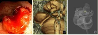
 We were recently asked by a colorectal surgeon to perform a virtual colonoscopy to localize a 2.5 cm tumor found on endoscopy and described as being " in the distal rectosigmoid". In this case virtual colonoscopy provided useful pre-op evaluation for the surgeon. The coronal cut 3D image and reconstruction simulating double contrast barium enema show the lesion in the distal sigmoid colon (yellow circle). Images obtained with Voxar Colonscreen software.
Click on Images for full screen full resolution

No comments:
Post a Comment